
DBS surgery.
DBS is implanted in two stages. Stage 1 places the leads in the brain, using either awake functional mapping or asleep real-time MRI. Stage 2 implants the pulse generator and connectors about two weeks later.
Stage 1, awake option: physiological mapping.
DBS lead implantation traditionally took place with patients awake, so that the implantation site could be optimized by functional brain mapping. Functional brain mapping involves recording from microelectrodes lowered into the brain before the DBS leads are inserted. These very thin electrodes record the activity of single neurons, which tells us when we are in the anatomic target. A robotic stereotactic assistant (ROSA) positions the trajectory guides and electrode holders, to ensure maximum implantation accuracy.
We also stimulate through these mapping electrodes to confirm the best trajectory for the DBS lead, based on thresholds for side effects and symptom improvement. Next, we place the DBS lead through the trajectory with the best mapping result and test that location with stimulation through the DBS lead. When we are happy with the result on the first side, we move to the second side. The patient is awake for mapping and stimulation testing, but typically asleep at the beginning and end of the procedure.
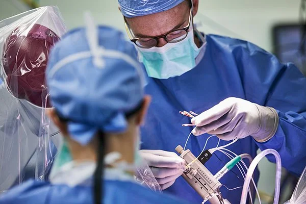
Dr. Richardson connecting the microelectrodes.

Dr. Herrington testing the effects of stimulation.
Stage 1, asleep option: real-time MRI.
Since 2010, we have implanted DBS leads using real-time MRI visualization with patients under general anesthesia. Instead of intraoperative microelectrode recording and stimulation testing, we verify accurate lead placement by direct anatomic visualization with intraoperative MRI. We use a targeting system called ClearPoint. Dr. Richardson helped launch this system during its preclinical development at the University of California San Francisco, and he established one of the earliest intraoperative MRI DBS programs at the University of Pittsburgh Medical Center in 2011. At MGH, this procedure is performed in an intraoperative 3-Tesla MRI suite.

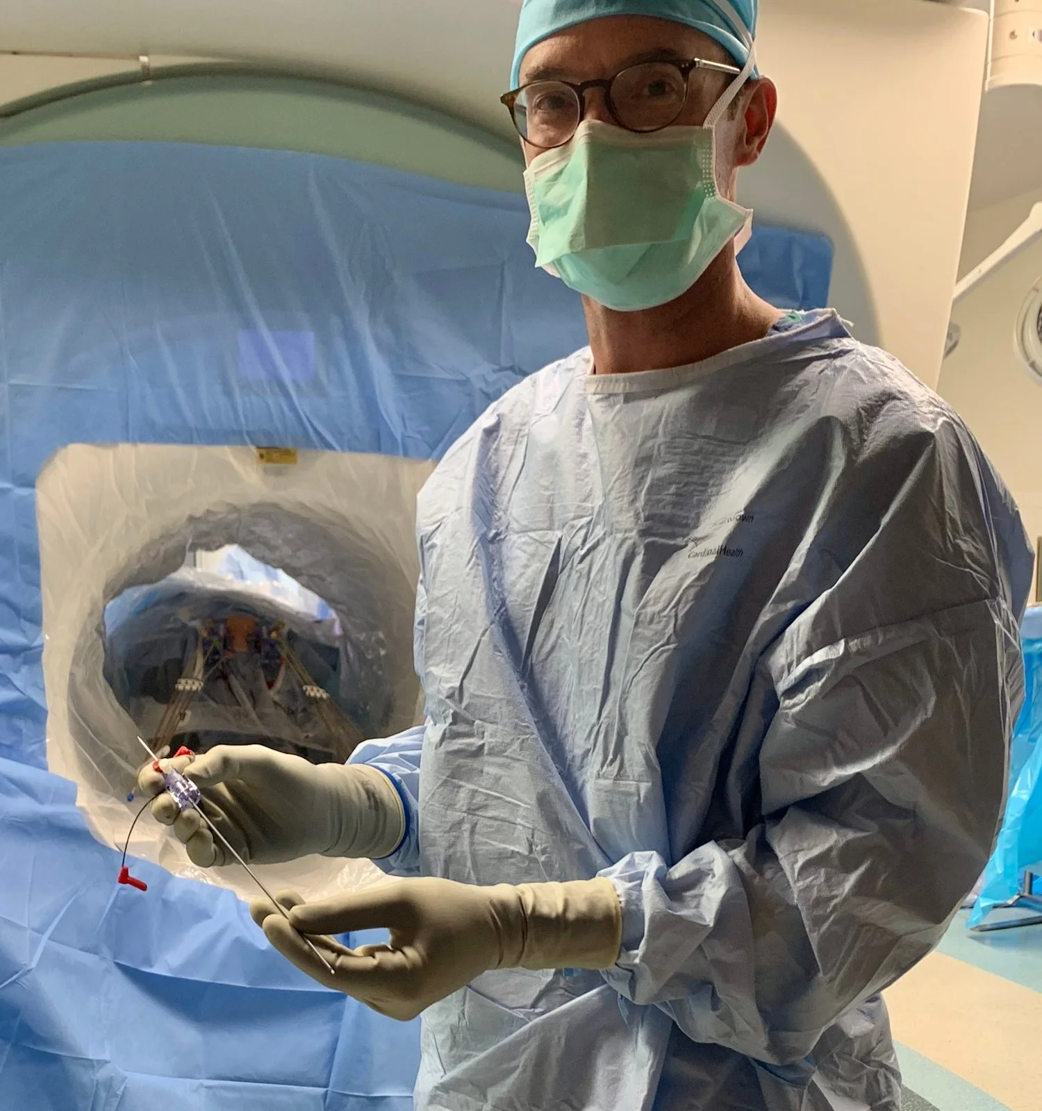
The ClearPoint system. Illustration of the SmartFrame and trajectory (left). Dr. Richardson in the intraoperative MRI operating room (right).
Video. Aspects of asleep iMRI-DBS at MGH. The surgery is performed within an intraoperative 3-Tesla MRI suite.
Awake versus asleep: which is best?
Patients undergoing DBS surgery are expected to do equally well, whether they have awake implantation with functional brain mapping or asleep implantation with real-time MRI visualization. Dr. Richardson and others have published case series showing that outcomes are similarly excellent with both options (read the article). Because we have extensive experience with both methods, we let patients choose the method they prefer. This chart may help you decide.
| Consideration | Awake | Asleep |
|---|---|---|
| Anesthesia for Stage 1 | Conscious sedation | General anesthesia |
| Anesthesia for Stage 2 | General anesthesia | General anesthesia |
| Target verification with real-time MRI | No | Yes |
| Target verification with functional mapping | Yes | No |
| Minimally invasive | Yes | Yes |
| Amount of hair shaved | Less | More |
| Patients with severe anxiety | No | Yes |
| Off medications for surgery | Yes | No |
| Patients with chronic neck pain | Not ideal | No problem |
| Children | Not ideal | No problem |
| Patients who want to participate in brain research | Possible | Possible |
Stage 2: implanting the pulse generator and connectors.
Stage 2 surgery connects the DBS leads by cables to the battery, or implantable pulse generator (IPG), which is placed in a skin pocket on the surface of the pectoralis muscle in the chest, just below the collarbone. The IPG is almost always placed on the right side of the chest, to reserve the left side for a cardiac pacemaker if one is ever needed.
Stage 2 is performed under general anesthesia for all patients and is typically separated from Stage 1 by about two weeks. The stages are separate because of the time required to perform both together, and to provide a window to evaluate lead placement with a postoperative CT scan. Every patient has a CT scan after Stage 1. The CT is fused to the preoperative MRI to model the position of the DBS leads. In fewer than 10% of cases, Dr. Richardson identifies an opportunity to change the depth of a DBS lead, which is easily done at Stage 2.
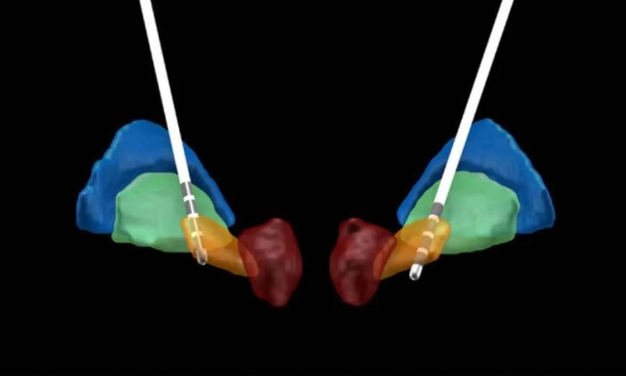
DBS lead reconstruction. Using special software, the post-operative CT is combined with the pre-operative MRI to generate an estimation of the lead locations within the target structure, for each patient. Here, the contacts of bilateral DBS leads are shown within the subthalamic nucleus (orange volume).